What Kind Of Tests Will Be Done During An Eye Exam?
Curious about the nature of tests carried out during an eye exam? This article informs you all about it. Imagine walking into either of the two locations of Fifty Dollar Eye Guy, a professional eye care establishment situated in Pensacola, FL. Here you encounter Dr. Joseph Tegenkamp whose friendliness and professionalism immediately puts you at ease. Their dedication to exceptional customer service and personal care of each patient is clearly evident, aiming for nothing less than a comfortable experience for you. Ultimately, they are intent on providing high-quality eye care which encompasses comprehensive eye exams adorned with a wide-selection of fashionable eyewear. This article will take you through the specific types of tests you can expect during an eye exam, putting your mind at ease for your next appointment.
Understanding the Purpose of an Eye Exam
Eye exams are a crucial aspect of maintaining overall health, much like regular doctor's visits or dental checks. Regular eye examinations ensure that your vision remains sharp and that any eye-related conditions or diseases are detected early.
The Importance of Regular Eye Examinations
Regular eye exams not only keep your prescription updated, ensuring clear vision, but are also essential in finding eye diseases early on. These screenings can often detect chronic systemic diseases like diabetes and high blood pressure. Remember, early detection gives you the best chance of successful treatment and preserves your vision.
General Objectives of an Eye Exam
The primary objective of an eye exam is to evaluate your eye health and determine the quality of your vision. During the exam, your eye doctor checks for common eyesight problems like nearsightedness, farsightedness, and astigmatism. They also look for eye diseases that could potentially harm your vision, such as glaucoma, cataracts, and macular degeneration.
Visual Acuity Test
Visual acuity tests are designed to measure the sharpness of your vision. This part of the eye examination checks how well you see the details of a letter or symbol from a distance.
Explanation of a Visual Acuity Test
During a visual acuity test, each of your eyes is individually tested for how well it can see. Your eye doctor will usually cover one eye and ask you to read letters or identify symbols of various sizes on a chart.
Tools Used in a Visual Acuity Test
Visual acuity tests usually involve the use of a standard eye chart, typically the Snellen chart. If you wear glasses or contacts, you'll be tested with them on.
How a Visual Acuity Test is Performed
During the test, you'll be asked to read lines of letters, each line becoming smaller as you progress. This determines the smallest line of letters you can correctly identify.
Interpreting Results of a Visual Acuity Test
Your visual acuity is expressed as a fraction, such as 20/20. The top number refers to the standard distance at which testing is performed (20 feet), and the bottom number indicates the smallest line size you were able to read. A 20/20 visual acuity means you can see at 20 feet what a person with normal vision can see at the same distance.
Peripheral Vision Test
Peripheral vision tests help identify blind spots (Scotomas), which could substantially affect your quality of life and indicate a wide range of potential eye health problems.
Importance of Testing Peripheral Vision
The peripheral vision test assesses the scope of your side vision (peripheral vision). This is particularly essential for daily activities such as driving, playing sports, even walking.
How a Peripheral Vision Test is Conducted
During the test, you'll cover one of your eyes and look straight ahead with the other, as the examiner moves an object slowly into your side vision to ascertain the extent of your peripheral vision.
Common Abnormalities Detected Through Peripheral Vision Test
Peripheral vision tests can detect glaucoma, retinitis pigmentosa and even certain conditions affecting the brain such as Parkinson's disease or strokes.
Color Blindness Test
Not being able to distinguish certain colors can significantly impact tasks associated with colors, from everyday tasks to activities at work.
Understanding Why Color Blindness Test is Conducted
Color blindness tests help your eye care professional determine if you have a color vision deficiency.
Procedure of a Color Blindness Test
A common method used for color blindness testing is the Ishihara Plate test. This consists of several colored plates, each containing a circle of dots appearing randomized in color and size. You would then identify a pattern within the dots differing in color.
What the Results of Color Blindness Tests Mean
Inability or difficulty in seeing the colored pattern signals potential color blindness aspects.
Ocular Motility Testing
Checking the movement of your eyes ensures they are working together to give you clear, comfortable vision.
Purpose of Ocular Motility Test
An ocular motility test checks how well your eyes follow a moving object and move between two separate fixed objects.
Procedure of an Ocular Motility Test
During the test, you might be asked to follow the movement of a handheld light with your eyes or quickly shift your focus between two objects located some distance apart from each other.
Common Conditions Diagnosed through Ocular Motility Testing
Problems detected during the ocular motility test may indicate conditions such as lazy eye, strabismus, or conditions that could lead to reading difficulties.
Depth Perception Test
Depth perception test allows your eye doctor to determine if you have an eye coordination problem that might affect how well you can judge the distance of objects.
What a Depth Perception Test is
The depth perception test assesses your ability to perceive objects in three dimensions (height, width, and depth) and the distance of an object.
How a Depth Perception Test is Done
Typically, this involves viewing and identifying 3D shapes or patterns on a chart.
Interpreting the Results of a Depth Perception Test
Problems in recognizing or judging depth or distance can suggest potential squint or lazy eye.
Cover Test
The primary function of a cover test is to check how well your eyes work together.
Overview of the Cover Test
A cover test is an examination that your doctor uses to determine if your eyes are aligning properly.
How a Cover Test is Performed
This exam is simple and straightforward. Your doctor will ask you to focus on a target at a distance and will cover each of your eyes alternately while you stare at the target. The way your eyes respond to the doctor's movements can indicate alignment problems.
Common Issues Diagnosed through a Cover Test
Conditions such as strabismus or binocular vision (where both eyes can't cooperate properly) that may lead to ocular discomfort, amblyopia (lazy eye) or even issues such as eyestrain and headaches, could be diagnosed through a cover test.
Retinoscopy
Retinoscopy is an exam your eye doctor uses to check your refractive error like nearsightedness, farsightedness, or astigmatism.
Understanding Retinoscopy
Retinoscopy involves your doctor observing the way light reflects from your eyes to estimate your prescription before fine-tuning it with further tests.
Procedure and Tools Used in Retinoscopy
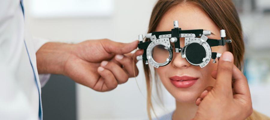
During a retinoscopy exam, your eye doctor dim the lights of the room and will ask you to focus on a large target (usually the big "E" on the eye chart). They'll then shine a light in your eyes and flip lenses in a machine in front of your eyes to estimate your prescription.
What Retinoscopy Can Reveal About Eye Health
Retinoscopy can accurately determine your refractive error – essential when prescribing corrective lenses. It can also detect issues with the clarity of your eyes' lens and other internal structures.
Tonometry (Eye Pressure Test)
Tonometry is used to measure the pressure within your eye, a critical step in screening and monitoring for glaucoma.
The Role of Tonometry in Eye Exams
Tonometry measures the fluid pressure inside the eye which is essential to keep the tissues healthy.
How Tonometry is Performed
During tonometry, a small amount of pressure is applied to the eye by a tiny device or a warm puff of air. The eye's resistance to the pressure allows the doctor to measure the internal pressure
What Elevated Eye Pressure Can Indicate
Increased eye pressure can indicate an enhanced risk of developing glaucoma. Tonometry is the most common method of detecting this risk before glaucoma can cause vision loss.
Dilation and Bio-microscope Exam
The pupillary dilation and bio-microscope exam give your eye doctor an in-depth view of the essential parts of your eye.
Understanding the Importance of Pupil Dilation in Eye Exams
Pupil dilation widens your eyes to allow more light to enter, helping your eye doctor see the retina and optic nerve more clearly.
Procedure of Dilation and Bio-microscope Exam
For the examination, your doctor will instill drops in your eyes to cause dilation, then use a bio-microscope (also called a slit lamp) to illuminate and magnify the structures of the eye.
Issues Often Detected Through a Dilation and Bio-microscope Exam
Through a dilation and bio-microscope examination, more serious conditions such as macular degeneration, retinal detachment, and even ocular tumors can be detected.


